If you’re experiencing disturbances in your gastrointestinal (GI) tract or your digestive system, your doctor may order an endoscopy, also called an esophagogastroduodenoscopy (EGD). Endoscopy is the gold standard for diagnosing most diseases and conditions of the upper GI tract. In some cases, endoscopy is also used to treat conditions as well. Read on to learn about the procedure as well as diseases detected by endoscopy.
‘The most exciting journey is the journey deep into the digestive tract……. exploring all those bends and curves, appreciating uniqueness of each surface and uncovering the secrets they hide is truly a rewarding experience.’ -Sandhya Shukla, MD
What Is an Endoscopy?
An upper GI endoscopy is used to diagnose and treat diseases or conditions of the upper digestive tract. The upper GI tract is considered to be from the mouth to the beginning of the small intestine (duodenum).
Certain symptoms may prompt you to call your doctor. Since diseases detected by endoscopy are many, the symptoms may vary widely. Many GI conditions share the same type of symptoms, so the best way to truly diagnose a GI problem is to get a much closer look. Symptoms that may warrant a call to your physician include:
- Abdominal pain
- Diarrhea, constipation, or both intermittently
- Nausea and vomiting
- Burning of the esophagus or stomach
- Regurgitation (food and liquid coming back up into the throat or mouth)
- Dysphagia (trouble swallowing)
- Noticeable changes in bowel habits
If you have symptoms such as these that persist and aren’t treated with conventional methods (such as prescribing proton pump inhibitors (PPIs) or antacids), you should let your health care provider know, so you can schedule the procedure.
Both the preparation for the endoscopy and the procedure itself are very straightforward. Your physician will likely advise you not to eat or drink anything after midnight, with the exception of sips of water to take medication.
A doctor performs an upper GI either in the office or hospital setting. It is an outpatient procedure that, from arrival to discharge, lasts under two hours. The endoscopy itself takes only about 10 minutes.
When you arrive, you’ll consult with the anesthesiologist, as you will be put under light anesthesia, the gastroenterologist, and the lead nurse. They will check your blood pressure, and give you an IV so you can receive the anesthesia.
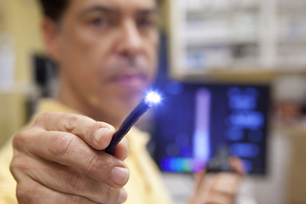
You are then taken to the procedure room and told to lie on your side. Anesthesia is administered, and the doctor performs the procedure. An endoscope is a long, thin flexible tube with a tiny camera attached at the end. Your physician will insert the endoscope through your mouth so it can take photographs of the entire GI tract, from the mouth to the duodenum (which is part of the small intestine).
Only so much can be seen with photographs, so it’s likely your doctor will take tissue samples and biopsies to be sent to the laboratory. Once the samples and biopsies return, your gastroenterologist can provide a more definitive diagnosis.
After the procedure, you are taken to a recovery room. The team will check for any side effects and monitor you for about half an hour. You must have someone to drive you home, but you can (in most cases) resume normal eating after the procedure. However, you should pause more physician activities for up to 24 hours.
Diseases and Conditions That Can Be Detected
Because many upper (and lower) GI conditions show similar signs, endoscopy is used so your gastroenterologist can provide you with the right diagnosis. There are many diseases detected by endoscopy; however, the more common ones are listed below.
Gastroesophageal Reflux Disease (GERD)
Gastroesophageal reflux disease (GERD) is very common. Its hallmark symptom is heartburn (acid reflux), although it may be accompanied by regurgitation. Reflux itself is due to stomach acid finding its way into the esophagus, where it doesn’t belong. There is a muscle at the bottom of the esophagus called the lower esophageal sphincter (LES). The LES should close completely after food or liquid passes through it, however, in some patients, the LES doesn’t work properly and can stay slightly or fully open. GERD is typically treated with medications (such as proton pump inhibitors) and lifestyle changes.
Helicobacter pylori (H. pylori)
Helicobacter pylori (H. pylori) infection is another common GI problem. It’s estimated that half the world has H. pylori present in their stomach, but for some, it causes inflammation of the stomach lining. Some patients are asymptomatic, while others experience symptoms such as abdominal pain and indigestion. H. pylori is a common cause of peptic (stomach) ulcers and can make you more predisposed to gastric cancer; however, stomach cancer is rare. H. pylori infection is typically treated with antibiotics to cure the infection and other medications (such as PPIs) to help with symptoms.
Esophageal Stricture and Dilatation
Those with esophageal stricture often have trouble swallowing, as esophageal stricture is the narrowing of the esophagus. This has many causes, but the most common cause is scarring as a result of acid reflux. More uncommonly, it can be caused by eosinophilic esophagitis (an “allergic” esophagus) or esophageal cancer.
Esophageal stricture can also be caused by esophageal dysmotility, which indicates a disorder in the way your esophagus moves. Stricture can often be detected by endoscopy, but often esophageal dilatation is used during the procedure to confirm the diagnosis. During esophageal dilatation, a balloon is placed in the stricture, which widens the esophagus. The balloon is then removed before the endoscopy is complete.
Stomach and Duodenal Ulcers
Stomach and duodenal ulcers are both considered peptic ulcers, which indicate a tear or break in the stomach lining. Peptic ulcers are most commonly caused by H. pylori, and the treatment includes antibiotics and acid-reducing medication to lessen symptoms if symptoms are present.
It was once thought that ulcers were triggered by stress, diet, or smoking, but the main causes are H. pylori infection or overuse of nonsteroidal anti-inflammatory drugs (NSAIDs).
Stomach Cancer & Esophageal Cancer
Gastric (stomach) cancer and esophageal cancer are both rare but are detectable on endoscopy. If cancer is present, treatment includes oncological procedures, such as radiation and chemotherapy. While gastric cancers are rare, symptoms can include abdominal pain, heartburn, trouble swallowing, weight loss, and nausea and vomiting, among others. As these symptoms are similar to other types of gastrointestinal distress, consult your doctor if these symptoms persist.
Barrett’s Esophagus
Barrett’s esophagus is a gastrointestinal condition caused by acid reflux damage and causes a change in the esophageal lower lining. Barrett’s esophagus is diagnosed during endoscopy and will likely require more aggressive treatment than GERD or acid reflux. This is because you are much more likely to develop esophageal cancer with this diagnosis.
The changes in the lower lining of the esophagus are often comorbid with dysplasia (pre-cancerous cells). Treatment for Barrett’s depends on whether there is no dysplasia, low-grade dysplasia, or high-grade dysplasia. If you have no dysplasia, your gastroenterologist will likely prescribe GERD medications (such as PPIs) and conduct regular endoscopies to monitor the cells in your stomach.
If you have low-grade dysplasia, your doctor may remove the damaged cells (endoscopic resection) during your endoscopy. Other treatments include radio-frequency ablation, which removes the damaged tissue, and cryotherapy, which is an endoscopic procedure where damaged cells are treated intermittently with cold, then heat, causing them to die (autophagy).
Liver Disease, Cirrhosis, Portal Hypertension
An endoscopy can detect many types of liver diseases as well, such as cirrhosis, esophageal varices, and portal hypertension. Liver diseases can include:
- Hepatitis
- Cirrhosis
- Liver cancer
- Fatty liver disease
- Nonalcoholic fatty liver disease (NAFLD)
Depending on the type of diagnosis, treatment may vary widely. Liver cancer is treated often with radiation, chemotherapy, or surgery.
Cirrhosis is the permanent scarring of the liver and a primary cause is alcohol use disorder (AUD). A precursor to cirrhosis is fatty liver, and typically the liver is a very elastic organ. A patient can recover from fatty liver or elevated liver enzymes by practicing abstinence from ethanol.
However, cirrhosis is often hard to treat because of the irreversible scarring—patients have a much better outcome if cirrhosis is caught in the early stages. The first-line treatment is always to stop drinking. NAFLD can also cause scarring of the liver and may require a liver transplant.
Often, gastroenterologists can diagnose liver disease through an endoscopy because portal hypertension is detected. Portal hypertension refers to elevated pressure within the portal venous system. Its main cause is cirrhosis (liver scarring).
Esophageal varices can also be detected via endoscopy, and they are a marker of liver disease. Varices are veins in the esophagus that can tear and bleed, which can be life-threatening. If esophageal varices are suspected, your physician may order an endoscopy as an emergency procedure, as this can treat active bleeding and esophageal varices.
Scheduling an Appointment Today
If you’re experiencing GI distress and upset that persists, schedule an appointment today, and our team of physicians and healthcare professionals will work together to help manage your symptoms, diagnose, and offer you quality and comprehensive treatment for all types of gastrointestinal disorders.
© All Rights Reserved


